博文
Detection MOAB enzyme and its oxidative stress by NIR probe
|
Ratiometric near-infrared fluorescent probe for synergistic detection of monoamine oxidase B and its contribution to oxidative stress in cell and mice aging models
Rui Wang【王蕊】,ab Xiaoyue Han【韩潇玥】,ab Jinmao You【尤进茂】,ab Fabiao Yu【于法标】*ab and Lingxin Chen【陈令新】*ab
a Key Laboratory of Life-Organic Analysis, Key Laboratory of Pharmaceutical Intermediates and Analysis of Natural Medicine, College of Chemistry and Chemical Engineering, Qufu Normal University, Qufu 273165, China.
b Key Laboratory of Coastal Environmental Processes and Ecological Remediation; Research Centre for Coastal Environmental Engineering and Technology, Yantai Institute of Coastal Zone Research, Chinese Academy of Sciences, Yantai 264003, China. fbyu@yic.ac.cn; lxchen@yic.ac.cn.
As new biomarkers, monoamine oxidases (MAOs) play important roles in maintaining the homeostasis of biogenic amines via catalyzing the oxidation of biogenic amines to corresponding aldehydes with the generation of reactive oxygen species (ROS). MAOs have two isoforms, MAO-A and MAO-B. MAO-A is considered to be a major factor of neuropsychiatric and depressive disorders. However, MAO-B is thought to be involved in several neurodegenerative diseases. Therefore, to explore their distinct roles in different diseases, the selective detection of MAOs is essential. Herein, two new types of NIR fluorescent probes, MitoCy-NH2 and MitoHCy-NH2, are provided for synergistic imaging of MAO-B and its contribution to oxidative stress in cells and in mice aging models. These probes are composed of three moieties: heptamethine cyanine as fluorophore, propanamide as recognition group, and triphenylphosphonium cation as mitochondrial targeting group. The amine oxidation and β-elimination reaction can lead to obvious fluorescence increase and color changes from green to blue. The probe MitoHCy-NH2 can be used to synergistically detect MAO-B and its contribution to oxidative stress in replicative senescence model. And the probe MitoCy-NH2 can offer ratiometric near-infrared fluorescence for the selective detection of MAO-B in H2O2-induced cell aging model and in mice aging models. The results reveal that there are different MAO-B levels in different age of mice models. MitoCy-NH2 also can evaluate therapeutic effects of pargyline and selegiline in mice models. The desirable analytical behaviors of our probes make they are useful chemical tools for the selective detection of MAO-B and its contribution to oxidative stress in biosystems.
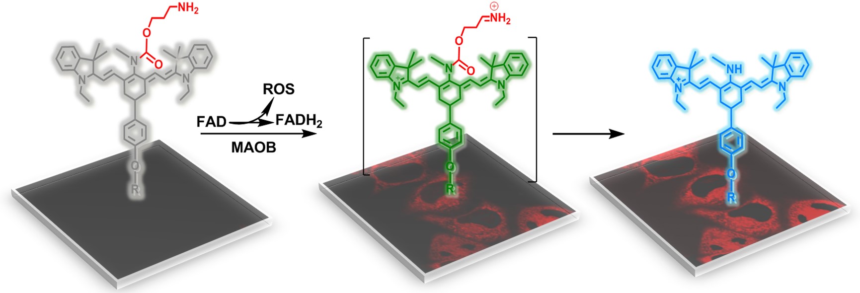
Scheme 1 Molecular structures and the proposed detection mechanism of MitoHCy-NH2.
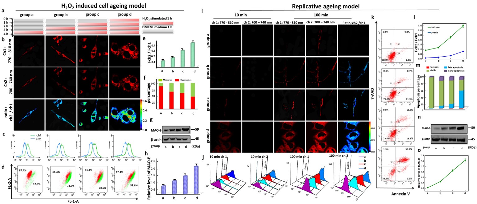
Figure 5 Synergistic imaging of MAO-B and O2•− in two types of cell ageing models. a) The protocol of H2O2 induced cell aging models: cells were divided into four groups and incubated with 550 μM H2O2 for 60 min, 120 min, 180 min, and 240 min, respectively. Then the cells were cultured for more two days; b) Fluorescent images with 5 μM MitoCy-NH2 for 100 min; Two fluorescence collection windows, channel 1:750 – 800 nm (λex = 730 nm); channel 2: 700 – 740 nm (λex = 650 nm). c) Flow cytometry analysis of the cells in b); d) Cell apoptosis analysis using JC-1 by analyzing mitochondrial membrane potentia in b); e) The average ratio values of ratiometric images in b); f) Statistical analysis of JC-1 in b). n = 5; g) Western blotting analysis of MAO-B in b); h ) Quantitative analysis of active MAO-B levels. n = 3. i) Replicative senescence cell aging models: cells were divided into 4 groups. Group a – d: the CPD of each group was 3, 15, 30, and 50. Fluorescent images with 5 μM MitoCy-NH2 at two time points: 10 min and 100 min. All the cells were incubated with5 μM MitoHCy-NH2 for 100 min, then washed with DMEM to remove the redundant probe. j) Flow cytometry analysis of the cells in i); k) Apoptosis analysis by Annexin V/ 7-AAD: (Q1) necrosis (AnnexinV-/7-AAD+), (Q2) late apoptosis (AnnexinV+/7-AAD+), (Q3) viable cells (AnnexinV-/7-AAD-), (Q4) early apoptosis (AnnexinV+/7-AAD-); l) The average ratio values in i); m) Apoptosis analysis of k); n) Western blotting analysis of MAO-B in i); o) Quantitative analysis of the levels of active MAO-B. n = 3.

Figure 6. Imaging of MAO-B levels in the mice brain with different ages. Fluorescence collection windows: channel 1: 760 - 840 nm, λex = 730 nm. Channel 2: 700 – 800 nm, λex = 650 nm. All the BALB/c mice were incubated MitoCy-NH2 (100 μM, 50 μL in 1:99 DMSO/ saline, v/v) for 30 min with intracranial injection. a) In vivo imaging of mice in group a – d, the age of the mice were 1, 6, 12, and 24-month-old, respectively; b) Ex vivo imaging of separated organs sacrificed from the mice in a); c) Imaging of fresh mice SNpc from b). Two fluorescence collection windows, channel 1: 750 – 800 nm (λex = 730 nm); channel 2: 700 – 740 nm (λex = 650 nm). d) and g) Western blotting analysis for MAO-B levels of mice in a). e) The average fluorescence ratios for a); f) The average fluorescence ratios for b).
Anal. Chem., 2018, 90 (6), pp 4054–4061
DOI: 10.1021/acs.analchem.7b05297
Publication Date (Web): February 5, 2018
https://pubs.acs.org/doi/abs/10.1021/acs.analchem.7b05297
https://blog.sciencenet.cn/blog-2438823-1124177.html
上一篇:Evaluation selenocysteine effect by ratiometric NIR probe
下一篇:Detection O2•- and Hg2+ via three-channel fluorescent probe
全部作者的其他最新博文
- • Triphenylamine-AIE Materials for Cancer Theranostics
- • Fluorescent Probe forRatiometric Monitoring of Peroxynitrite
- • Fluorescence Probe for Pathological Stages of Wound Healing
- • Macrophage M2 polarization to neurological damage
- • SERS-RCA biosensor for profiling dual miRNAs
- • a glutathione-activated near-infrared fluorescent probe