博文
Mitochondria-targeting probe for imaging of cysteine
|
Mitochondria-targeting near-infrared ratiometric fluorescent probe for selective imaging of cysteine in orthotopic lung cancer mice
Xiaoyu Zhang【张晓玉】,a,b,d Na He【赫娜】,a,b,d Fabiao Yu【于法标】,c,d* Lingxin Chen【陈令新】b,d* and Changjun Lv【吕长俊】a,d*
a Department of Respiratory Medicine, Binzhou Medical University Hospital, Binzhou 256603, China
b Key Laboratory of Coastal Environmental Processes and Ecological Remediation, Yantai Institute of Coastal Zone Research, Chinese Academy of Sciences, Yantai 264003, China
c Institute of Functional Materials and Molecular Imaging, College of Clinical Medicine, Key Laboratory of Hainan Trauma and Disaster Rescue, College of Emergency and Trauma, Hainan Medical University, Haikou 571199, China
d Medicine Research Center, Institute of Molecular Medicine, Binzhou Medical University, Yantai 264003, China
Corresponding Author E-mail: fbyu@yic.ac.cn (F. Yu), lxchen@yic.ac.cn (L. Chen), lucky_lcj@sina.com (C. Lv)
Abstract: Cysteine (Cys) plays significant roles in many physiological processes, although its normal concentration is maintained at the micromole level. Abnormally high levels of intracellular Cys can lead to many diseases including cancer. Recent years, many effective fluorescent probes have been developed for the selective detection of Cys against other biological thiols. Herein, we synthesized a ratiometric near-infrared (NIR) fluorescent probe Cy-OAcr for selective imaging of intracellular Cys. Cy-OAcr has a lipophilic iminium cation unit as the mitochondrial guider and an acrylate group as the Cys recognition unit as well as a fluorescence modulator for rearranging the conjugated π-electron system of cyanine fluorophore. Upon detection of Cys, there occurs a significant absorption and fluorescence spectral shift, which are desirably beneficial for ratiometric detection. This probe has high sensitivity and selectivity for Cys detection over glutathione (GSH), homocysteine (Hcy), and other biomolecules with a low limit of detection at 0.09 μM. Probe Cy-OAcr is capable to detect and image Cys in three living cancer cell lines and their corresponding tumor-bearing mice models. More importantly, we successfully apply this fluorescent probe to evaluate the level of Cys in orthotopic lung cancer model. Imaging analyses reveal that the probe can discriminate tumor lesions from normal tissues, indicating its significant potential applications for clinical auxiliary diagnosis of cancer.
Key words: Fluorescent probes; Near-infrared fluorescence; Cysteine; Mitochondria target; Lung cancer
https://www.sciencedirect.com/science/article/pii/S0925400518320100
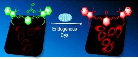
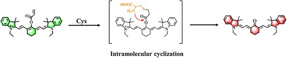
Scheme 2. Molecular structure and proposed mechanism of Cy-OAcr for the ratiometric detection of Cys.
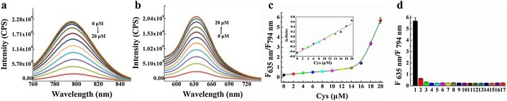
Fig. 1. Spectral properties and selectivity of Cy-OAcr. a) Dose-dependent emission spectra (λex = 750 nm) of Cy-OAcr (10 μM) towards Cys. b) Dose-dependent emission spectra (λex = 535 nm) of Cy-OAcr(10 μM) towards Cys. c) The fluorescence ratio signal (FRatio = F635 nm/F794 nm) of Cy-OAcr towards Cys. Inset: the linear relationship between Lg(Ratio) and Cys. d) Selectivity of Cy-OAcr towards different biologically reactive species. 1, Cys (0.2 mM); 2, GSH (8 mM); 3, Hcy (0.2 mM); 4, H2S (0.1 mM); 5, Asp (1 mM); 6, Glu (1 mM); 7, Gly (1 mM); 8, Lys (1 mM); 9, BSA (1 mM); 10, NO (0.1 mM); 11, H2O2 (0.1 mM); 12, HOCl (0.1 mM); 13, Ca2+ (1 mM); 14, Fe2+ (0.2 mM); 15, Zn2+ (0.2 mM); 16, Br− (0.1 mM); 17, Cl− (0.1 mM). Date were recorded at 45 min. The data were shown as mean (±s.d.) (n = 7).
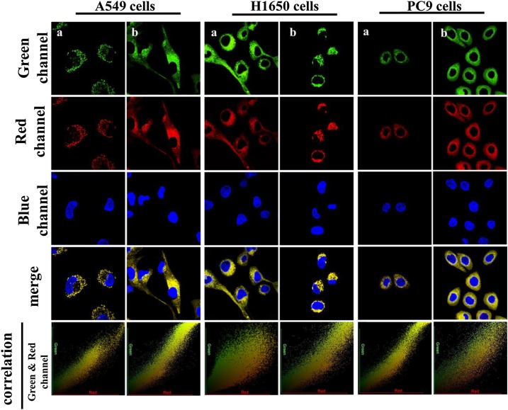
Fig. 2. Mitochondrial multicolor colocalization of Cy-OAcr (Red channel) with MitoTracker Green (Green channel a), Rhodamine 123 (Green channel b) and Hoechst 33,342 (Blue channel) in A549 cell line, H1650 cell line and PC9 cell line. The cells were incubated with probe Cy-OAcr (10 μM) for 30 min, Mito-Tracker Green (1 μg mL−1) for 15 min, Rhodamine 123 (1 μg mL−1) for 15 min, and nuclear dye Hoechst 33,342 (1 μg mL−1) for 30 min, respectively. Green channel a: λex = 488 nm, λem = 450–550 nm. Green channel b: λex = 515 nm, λem = 550–600 nm. Red channel: λex = 550 nm, λem = 610–700 nm. Blue channel: λex = 405 nm, λem = (For interpretation of the references to colour in this figure legend, the reader is referred to the web version of this article).410–510 nm.
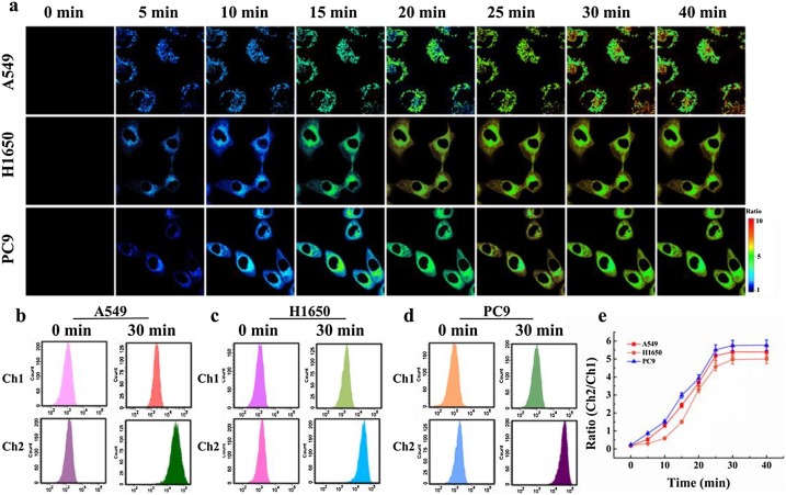
Fig. 3. Real-time ratio images and flow cytometry analysis endogenous Cys. a) Pseudocolor ratio images of endogenous Cys levels in A549, H1650 and PC9 cells at time points: 0, 5, 10, 15, 20, 25, 30 and 40 min. Fluorescence images collection windows for Channel 1: λex = 635 nm, λem = 700–800 nm. Channel 2: λex = 550 nm, λem = 600–700 nm. b) – d) Flow cytometry analysis of a). Channel 1: λex = 633 nm, λem= 750–810 nm. Channel 2: λex = 488 nm, λem = 610–670 nm. e) Plots of average ratio intensities of a). Ratios in e) were calculated through the fluorescence intensity ratio from Ch2/Ch1. The data were shown as mean (±s.d.) (n = 7).
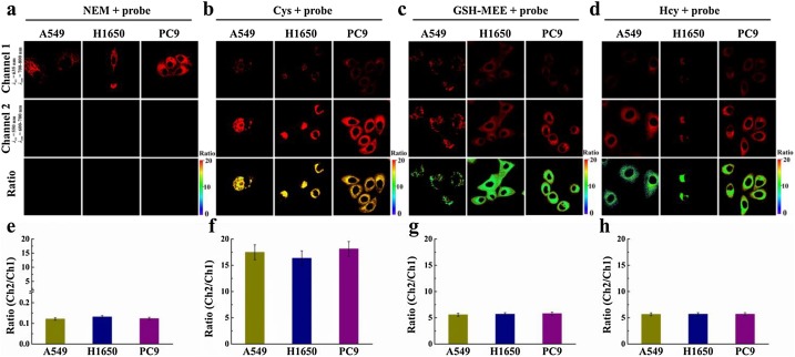
Fig. 4. Ratio images of the Cys in A549, H1650 and PC9 cells exposed to different stimulation agents. a) - d) The cells were pretreated with NEM (300 μM), Cys (200 μM), GSH-MEE (5 mM) and Hcy (200 μM) for 30 min and then incubated with Cy-OAcr (10 μM) for 30 min, respectively. Fluorescence images collection windows: Channel 1: λex = 635 nm, λem = 700–800 nm. Channel 2: λex = 550 nm, λem = 600–700 nm. e) - h) The average ratio fluorescence intensity of a) - d). Ratios in e) – h) were calculated through the fluorescence intensity ratio from Ch2/Ch1. The data were shown as mean (±s.d.) (n = 7).
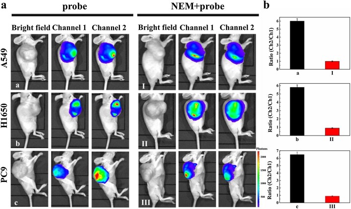
Fig. 5. In vivo imaging Cys in A549, H1650 and PC9 tumor-bearing mice. a) Group a, Group b and Group c were intratumorally injected Cy-OAcr (10 μM, 200 μl, in DMSO : saline = 1 : 99, v/v) for 1 h and then for in vivo imaging. Group I, Group II and Group III were intratumorally preinjected NEM (10 mM, 200 μL) for 1 h, subsequently injection of Cy-OAcr (10 μM, 200 μl, in DMSO : saline = 1 : 99, v/v) for 1 h, and then for in vivo imaging. The total number of photons from the entire tumor lesions of the mice were integrated. b) The average ratio intensity value of a). Ratios in b) were calculated through the mean number of photons ratio from Ch2/Ch1. The data were shown as mean (±s.d.) (n = 7).
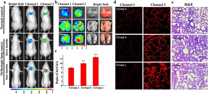
Fig. 6. In vivo imaging Cys in orthotopic lung cancer mice. a) In vivo fluorescent imaging. Fluorescence collection windows: Channel 1: λex = 740 nm, λem = 750–830 nm. Channel 2: λex = 520 nm, λem = 580–660 nm. b) Isolated lung fluorescence imaging. c) The average ratio intensity value of Group a–c at 30 min. Ratios in c) were calculated through the mean number of photons ratio from Ch2/Ch1. Difference was analyzed by one-way ANOVA and LSD post-test. ***P < 0.001 vs Group a. d) Confocal imaging of fresh lung tissue slice of Group a, b and c. Fluorescence images collection windows for Channel 1: λex = 635 nm, λem = 700–800 nm. Channel 2: λex = 550 nm, λem = 600–700 nm. Scale bar = 10 μm. e) H&E stained lung tissue from normal and cancer lesions in Group a–c. Scale bar = 20 μm. The data were shown as mean (±s.d.) (n = 7).
Xiaoyu Zhang is currently a physician at Yantai Affiliated Hospital of Binzhou Medical University. She received her master candidate, joint-educated, at Binzhou Medical University, and Yantai Institute of Coastal Zone Research, Chinese Academy of Sciences, during 2015 - 2018. She received her BS from Binzhou Medical University, in 2015. Her current research interest is focused on the accurate diagnosis of pulmonary fibrosis and lung cancer.
Fabiao Yu is currently a professor at Institute of Functional Materials and Molecular Imaging, Hainan Medical University. He served for Yantai Institute of Coastal Zone Research, Chinese Academy of Sciences during 2013 - 2017. He received his PhD, joint-educated, at Dalian University of Technology, and Dalian Institute of Chemical Physics, Chinese Academy of Sciences, in 2013. His research interests focus on functional probe molecules (fluorescence and phosphorescence analysis), theranostics, and organic functional materials.
Changjun Lv is now a vice president in Binzhou Medical University. He is a professor and serves in Affiliated Hospital of Binzhou Medical University as a chief physician. He received his MD from China Medical University in 1995. He is skilled in the diagnosis and treatment of respiratory diseases, especially in the diagnosis of interstitial lung diseases. He is an expert with China’s State Council special allowance due to his excellent performance in clinical work. His research interests include diagnosis and therapy of idiopathic pulmonary fibrosis, drug developments, and theranostics.
https://blog.sciencenet.cn/blog-2438823-1147085.html
上一篇:从你的全世界路过: 猪头的爱情----我的大学
下一篇:Imaging analysis of HNO anti-inflammatory effects
全部作者的其他最新博文
- • Triphenylamine-AIE Materials for Cancer Theranostics
- • Fluorescent Probe forRatiometric Monitoring of Peroxynitrite
- • Fluorescence Probe for Pathological Stages of Wound Healing
- • Macrophage M2 polarization to neurological damage
- • SERS-RCA biosensor for profiling dual miRNAs
- • a glutathione-activated near-infrared fluorescent probe