博文
NIR ratiometic fluorescent probe for mercury exposure
|
A sulfydryl-based near-infrared ratiometic fluorescent probe for assessment of acute/chronic mercury exposure via associated determination of superoxide anion and mercury ion in cells and in vivo
Yue Wang【王悦】 Min Gao【高敏】 Chunyang Liao【廖春阳】 FabiaoYu【于法标】* LingxinChen【陈令新】*
aCAS Key Laboratory of Coastal Environmental Processes and Ecological Remediation, Research Centre for Coastal Environmental Engineering and Technology, Yantai Institute of Coastal Zone Research, Chinese Academy of Sciences, Yantai, 264003, China
bState Key Laboratory of Environmental Chemistry and Ecotoxicology, Research Center for Eco-Environmental Sciences, Chinese Academy of Sciences, Beijing, 100085, China
cUniversity of Chinese Academy of Sciences, Beijing, 100049, China
Received 22 March 2019, Revised 21 August 2019, Accepted 23 August 2019, Available online 26 August 2019.
Sensors and Actuators B: Chemical
Available online 26 August 2019, 127038
https://doi.org/10.1016/j.snb.2019.127038
Highlights
•The fluorescent probe HCy-SH for efficient associated detection of O2•-and Hg2+ with high selectivity and sensitivity.
•Compare to chronic mercury exposure, O2•- burst is much stronger in acute mercury exposure especially in the heart.
•Hg2+ mainly accumulated in kidney weather in acute mercury poisoning or chronic mercury poisoning.
Abstract
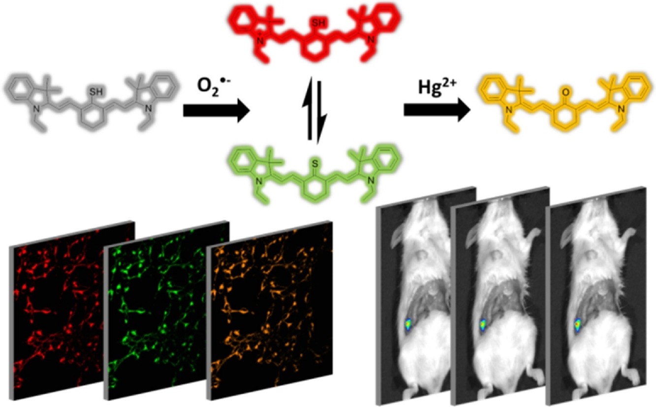
Mercury exposure is associated with severe oxidative stress especially superoxide anion radicals (O2•-). Mercury exposure is common in the clinic, but it is difficult to track. Although many researchers have tried to reveal the mechanism of mercury exposure, it is still remain vague that the differences between acute mercury exposure and chronic mercury exposure as well as the fluctuations in O2•- during mercury ion (Hg2+) stress. Thus effective tool for O2•- and Hg2+ associated-detection is needed urgently. Herein we have developed a stable near-infrared ratiometric fluorescent probe, HCy-SH, for O2•- and Hg2+ associated-detection. Probe HCy-SH was designed and synthesized based on a heptamethine cyanine fluorophore and a thiol-responsive group. The probe HCy-SH can be used for mercury poisoning detection in HEK 293 cells and mice models with low detection limits of 65 nM for O2•- and 72 nM for Hg2+. Relying on the probe HCy-SH, we found that O2•- burst was much severer in acute mercury exposure than chronic mercury exposure, especially in heart, and Hg2+ mainly accumulated in kidney no matter acute mercury exposure or chronic mercury exposure. The experimental results indicated that the probe HCy-SH was a potential candidate for accurate diagnosis and efficacy evaluation of mercury exposure.
Keywords
Mercury poisoning
Near-infrared fluorescent probe
Ratiometric biomaging
Superoxide anion
Mercury ion
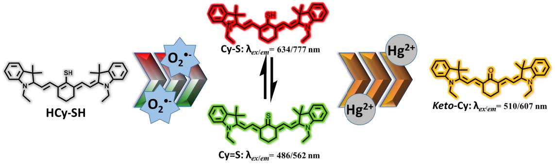
Scheme 1: Proposed associated-detection mechanism of HCy-SH for O2•- and Hg2+
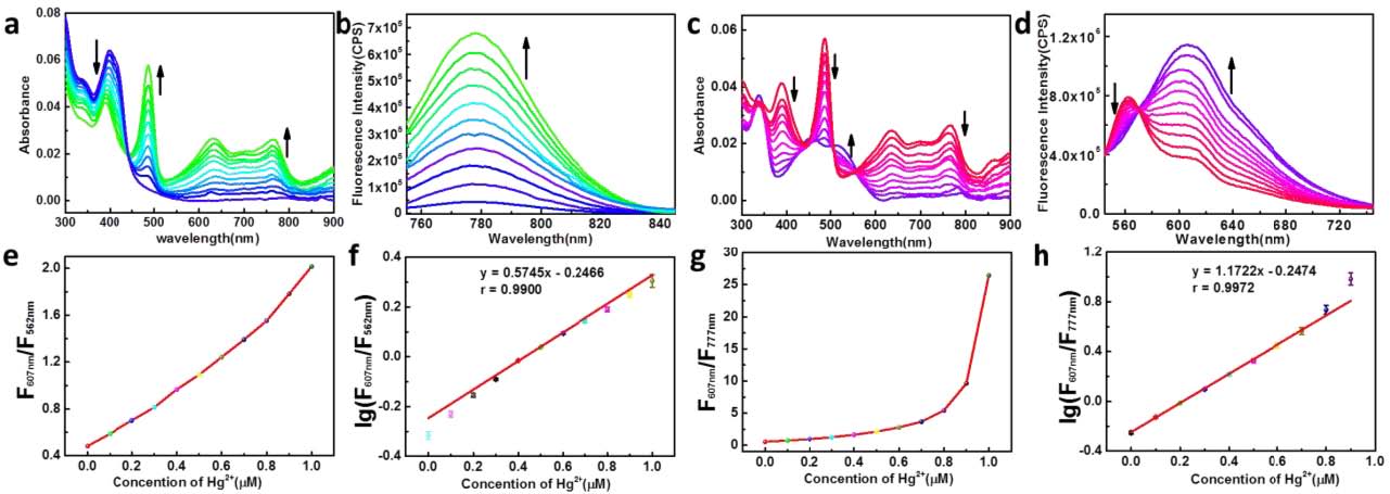
Figure 1. Spectra properties of the probe HCy-SH. Absorption (a) and emission (b) spectra of HCy SH (1μM) towards O2•- (0-1μM). Absorption (c) and emission (d) spectra of HCy-SH (1μM) + O2
•- (1μM) towards Hg2+ (0-1μM).Ratio signals (F607nm/F562nm) (e) and (F607nm/F777nm) (g) toward Hg2+ concentration (0-1μM). The corresponding linear relationships between the lg (F607nm/F562nm) (f) / lg(F607nm/F777nm) (h) and the Hg2+ concentration (0-1μM).
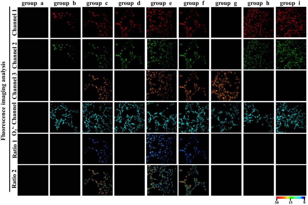
Figure 2. O2•- and Hg2+ detection in HEK 293 cells using laser scanning confocal microscope. Channel 1 λem = 755 -840 nm (λex = 635 nm); channel 2 λem = 545 - 570 nm (λex = 488 nm); channel 3 λem = 570 - 670 nm (λex = 535 nm).Ratio 1: channel 3 vs channel 2; Ratio 2: channel 3 vs channel 1. The commercial dye channel (O2•- channel) from 500to 530 nm (λex = 488 nm). group a: 1 μM HCy-SH; group b: 1 μM HCy-SH, 1 μM O2•-; group c: 1 μM HCy-SH, 1 μMO2•-, 1 μM Hg2+; group d: 1 μM HCy-SH, 10 nM PMA; group e: 1 μM HCy-SH, 10 nM PMA, 1 μM Hg2+; group f:
1μM HCy-SH, 1 μM Hg2+; group g: 1μM HCy-SH, 6 μM Hg2+; group h: 1μM HCy-SH, 3 μM sodium selenite, 1 μMHg2+; group i: 1μM HCy-SH, 3 μM selenocysteine, 1 μM Hg2+.
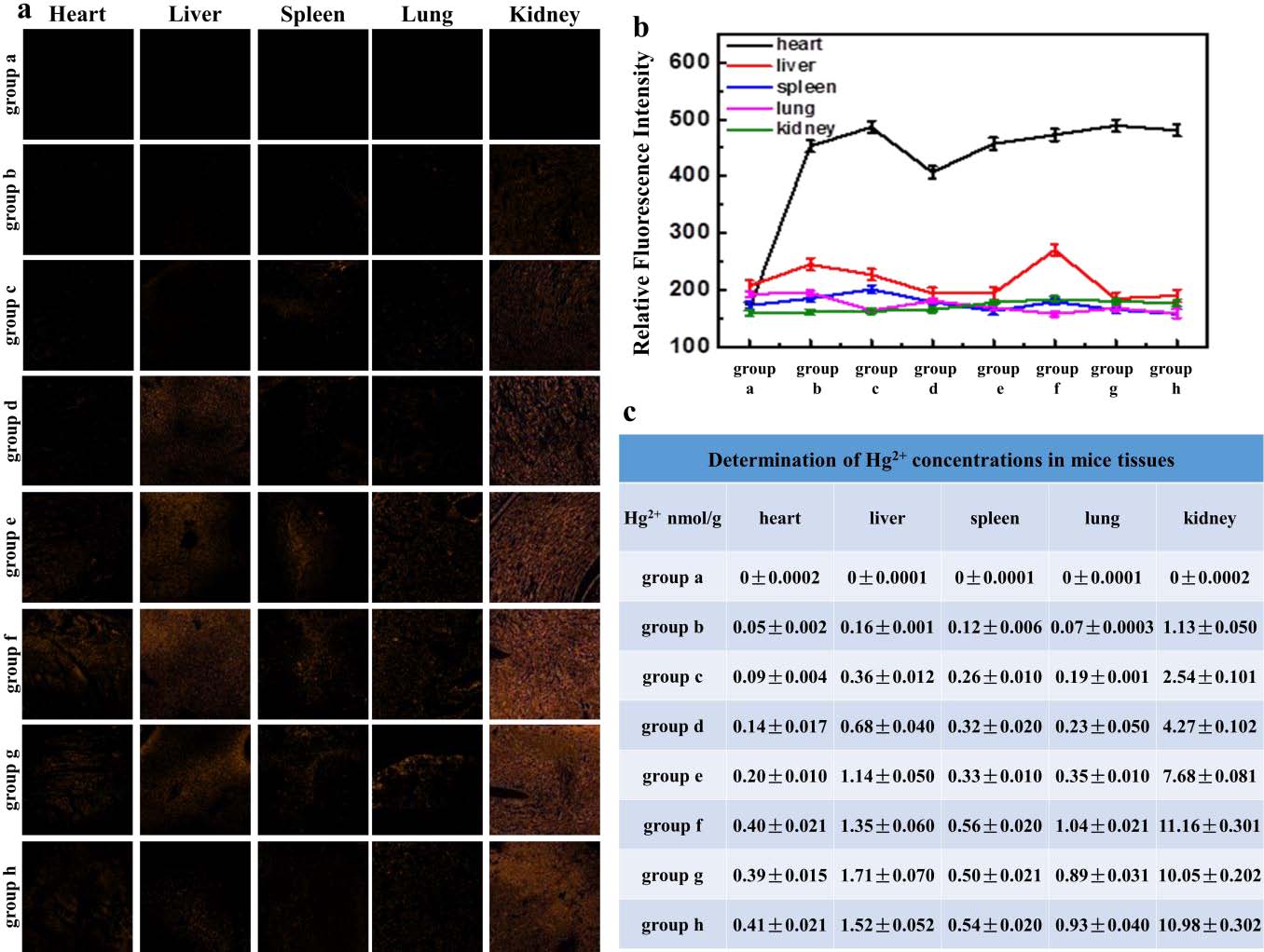
Figure 3. O2•- and Hg2+ associated-detection in organ slices from model mice. (a) Hg2+ detection in organ slices from model mice. (b) O2•- detection in different organs through tissue homogenate with ROS probe. (c) Hg2+ detection in different organs by ICP-MS. group a: 0.2 ml saline; group b: 4 mg/kg HgCl2, 0.2 ml; group c: 8 mg/kg HgCl2, 0.2 ml; group d: 12 mg/kg HgCl2, 0.2 ml; group e: 16 mg/kg HgCl2, 0.2 ml; group f: 20 mg/kg HgCl2, 0.2 ml; group g: 20 mg/kg HgCl2, 0.2 ml. 2.89 μM/kg sodium selenite, 0.2 ml; group h: 20 mg/kg HgCl2, 0.2 ml. 2.89 μM/kg selenocysteine, 0.2 ml.
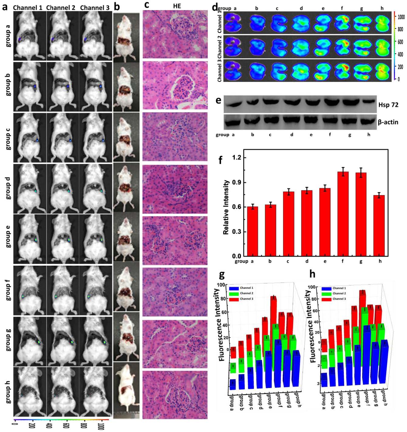
Figure 4. O2•- and Hg2+ associated-detection in mice model of acute mercury exposure. (a) Orthotopic injection assay.(b) Bright fields of the mice in (a). (c) H&E staining of the kidney sections (d) Ex vivo fluorescence imaging of separated kidneys from the mice in different groups. (e) Western blot analysis of hsp 72. (f) The relative intensity analysis of hsp 72. (g) The relative fluorescence quantitative analysis of (a). (h) The relative fluorescence quantitative analysis of (d). group a: 0.2 ml saline; group b: 4 mg/kg HgCl2, 0.2ml; group c: 8 mg/kg HgCl2, 0.2ml; group d: 12 mg/kg HgCl2, 0.2ml; group e: 16 mg/kg HgCl2, 0.2ml; group f: 20 mg/kg HgCl2, 0.2ml; group g: 20 mg/kg HgCl2,
0.2ml. 2.89 μM/kg sodium selenite; group h: 20 mg/kg HgCl2, 0.2ml. 2.89 μM/kg selenocysteine.
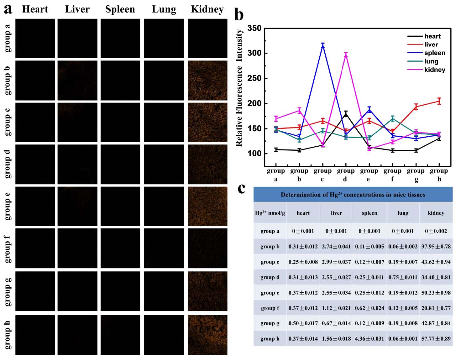
Figure 5. (a) Hg2+ detection in different organ slices. (b) O2•- detection in different organs with commercial ROS probe. (c) Hg2+ detection in different organs by ICP-MS. group a: 0.2 ml saline; group b: 18 mg/kg HgCl2 for 10 days; group c: 18 mg/kg HgCl2 for 15 days; group d: 18 mg/kg HgCl2 for 20 days; group e: 18 mg/kg HgCl2 for 25 days; group f: 18 mg/kg HgCl2 for 30 days; group g: 18 mg/kg HgCl2 for 30 days and 2.89 μM/kg sodium selenite for 30 days; group h: 18 mg/kg HgCl2 for 30 days and 2.89 μM/kg selenocysteine for 30 days
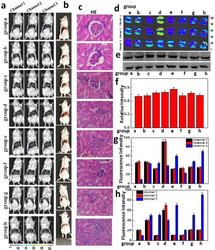
Figure 6. O2•- and Hg2+ associated-detection in mice model of chronic mercury exposure. (a) Orthotopic injection assay. (b) Bright fields of the mice in (a). (c) H&E staining of the kidney sections. (d) Ex vivo fluorescence imaging of separated kidneys from the mice in (a). (e) Western blot analysis of hsp 72. (f) The relative intensity analysis of hsp 72. (g) The relative fluorescence quantitative analysis of (a). (h) The relative fluorescence quantitative analysis of (d). group a: 0.2 ml saline; group b: 18 mg/kg HgCl2 for 10 days; group c: 18 mg/kg HgCl2 for 15 days; group d: 18 mg/kg HgCl2 for 20 days; group e: 18 mg/kg HgCl2 for 25 days; group f: 18 mg/kg HgCl2 for 30 days; group g: 18
mg/kg HgCl2 for 30 days and 2.89 μM/kg sodium selenite for 30 days; group h: 18 mg/kg HgCl2 for 30 days and 2.89 μM/kg selenocysteine for 30 days.
https://blog.sciencenet.cn/blog-2438823-1195914.html
上一篇:[转载]句型与结构两不误——从句型分类看论文结构
下一篇:SERS-based immunoassay for dual cardiac biomarkers detection
全部作者的其他最新博文
- • Triphenylamine-AIE Materials for Cancer Theranostics
- • Fluorescent Probe forRatiometric Monitoring of Peroxynitrite
- • Fluorescence Probe for Pathological Stages of Wound Healing
- • Macrophage M2 polarization to neurological damage
- • SERS-RCA biosensor for profiling dual miRNAs
- • a glutathione-activated near-infrared fluorescent probe