博文
Naphthorhodafluor fluorescent probe for hydrogen polysulfide
|
A semi-naphthorhodafluor-based red-emitting fluorescent probe for tracking of hydrogen polysulfide in living cells and zebrafish
YingyingMa[马莹莹]Zhencai Xu[徐振彩]QiSunLinlinWang【王琳琳】HengLiu【刘恒】*FabiaoYu【于法标】*
aMinistry of Education Key Laboratory for the Synthesis and Application of Organic Functional Molecules, College of Chemistry and Chemical Engineering, Hubei University, Wuhan 430062, China
bKey Laboratory of Emergency and Trauma, Ministry of Education, Key Laboratory of Hainan Trauma and Disaster Rescue, The First Affiliated Hospital of Hainan Medical University, Institute of Functional Materials and Molecular Imaging, College of Emergency and Trauma, Hainan Medical University, Haikou 571199, China
cKey Laboratory for Green Chemical Process of Ministry of Education and School of Chemistry and Environmental Engineering, Wuhan Institute of Technology, Wuhan 430205, China
Spectrochimica Acta Part A: Molecular and Biomolecular Spectroscopy
Volume 247, 15 February 2021, 119105
https://www.sciencedirect.com/science/article/pii/S1386142520310842
Highlights
•A semi-naphthorhodafluor-based red-emitting fluorescent probe SNARF-H2Sn for selective detection of H2Sn was designed.
•The addition of H2Sn would result in a > 1000-fold fluorescence enhancement within 10 min.
•SNARF-H2Sn was successfully employed to image exogenous/endogenous H2Sn in living cells and zebrafish.
Abstract
Hydrogen polysulfides (H2Sn, n ≥ 2) is recently regarded as a potential signaling molecule which shows a higher efficiency than hydrogen sulfides (H2S) in regulating enzymes and ion channels. However, the development of specific fluorescent probes for H2Sn with long-wavelength emission (>600 nm) are still rare. In this work, a semi-naphthorhodafluor-based red-emitting fluorescent probe SNARF-H2Sn containing a phenyl 2-(benzoylthio) benzoate responsive unit was constructed. SNARF-H2Sn was capable of selectively detecting H2Sn over other reactive sulfur species. Treatment with H2Sn would result in a > 1000-fold fluorescence enhancement within 10 min. SNARF-H2Sn showed a low limit of detection down to 6.7 nM, and further enabled to visualize exogenous/endogenous H2Sn in living A549 cells and zebrafish.
Graphical abstract

Keywords
Fluorescent probes Hydrogen polysulfide Red-emitting Cell imaging In vivo imaging
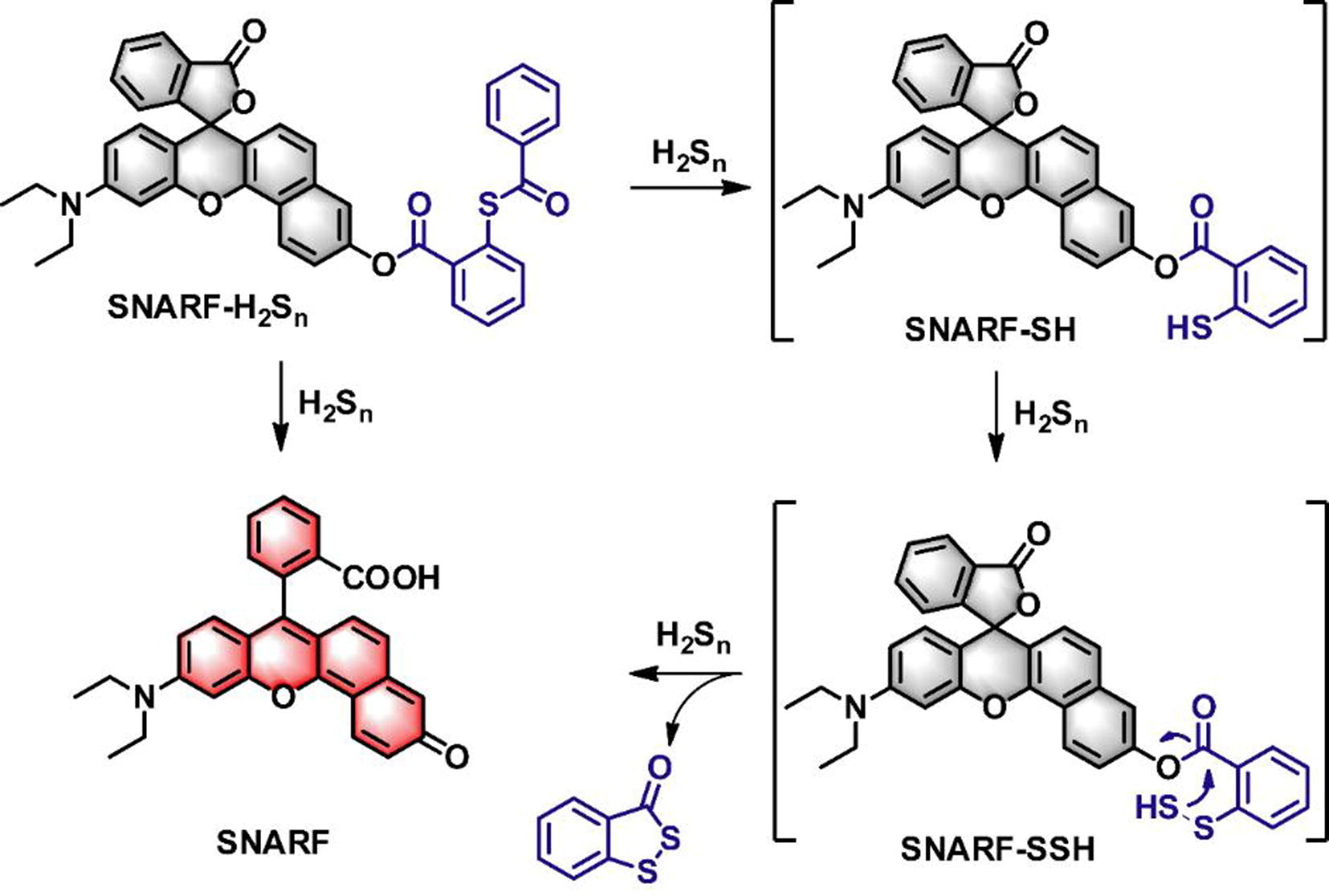
Scheme 1. Reaction mechanism of SNARF-H2Sn toward H2Sn.
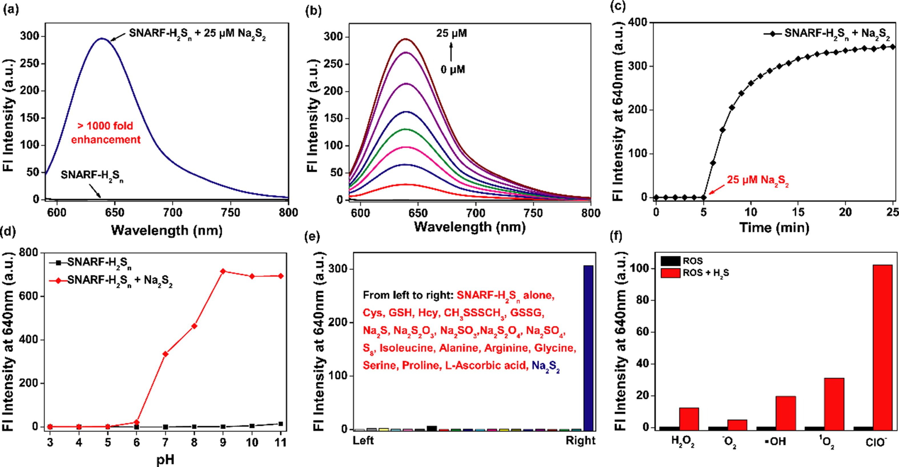
Fig. 1. (a) Fluorescence spectra of SNARF-H2Sn (10 μM) in the presence and absence of Na2S2 (25 μM). (b) Fluorescence responses of SNARF-H2Sn (10 μM) upon addition of Na2S2 (0–25 μM) for 10 min. (c) Time courses of fluorescence intensities at 640 nm before and after addition of Na2S2 (25 μM). (d) Influence of pH on the response of SNARF-H2Sn (10 μM) in the presence and absence of Na2S2 (25 μM). (e) Fluorescence responses of SNARF-H2Sn (10 μM) at 640 nm toward various potential biological analytes. From left to right: blank, Cys (200 μM), GSH (1 mM), Hcy (200 μM), CH3SSSCH3 (100 μM), GSSG (100 μM), Na2S (100 μM), Na2S2O3 (100 μM), Na2SO3 (100 μM), Na2SO4 (100 μM), S8 (50 μM), Ile (100 μM), Ala (100 μM), Arg (100 μM), Gly (100 μM), Ser (100 μM), Pro (100 μM), l-ascorbic acid (100 μM), Na2S2 (25 μM). (f) Fluorescence responses of SNARF-H2Sn (10 μM) at 640 nm in the presence of ROS (with or without 100 μM Na2S). From left to right: H2O2 (200 μM), (2) O2− (100 μM),  OH (100 μM), 1O2 (100 μM) and ClO− (100 μM).
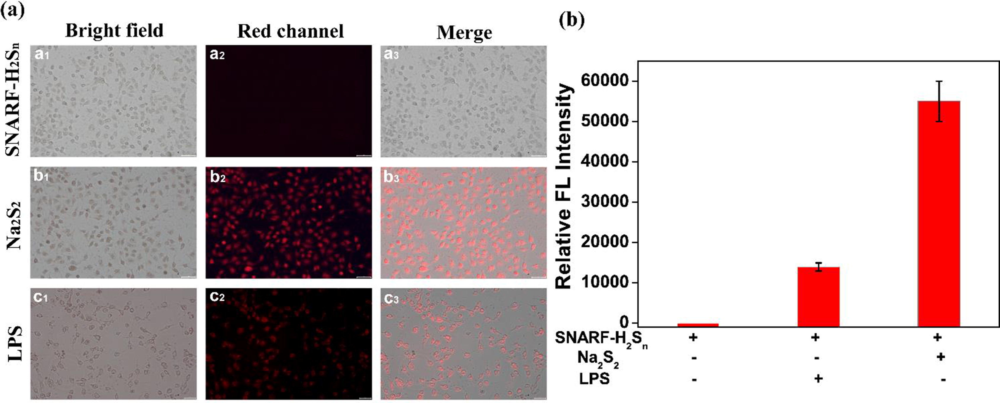
Fig. 2. (a) Fluorescence imaging of exogenous/endogenous H2Sn in living A549 cells. (a1–a3) The cells were incubated with 10 μM SNARF-H2Sn for 30 min; (b1–b3) the cells stained with 50 μM Na2S2 for 30 min were treated with 10 μM SNARF-H2Sn for another 30 min; (c1–c3) the cells stimulated with LPS for 12 h, and then incubated with 10 μM SNARF-H2Sn for 30 min. (a1, b1, c1) bright field images; (a2, b2, c2) red channel images; (a3, b3, c3) merged images. Scale bar = 50 μM. (b) Relative fluorescence intensity of red channel in panel A. Values represent mean standard error (n = 3).
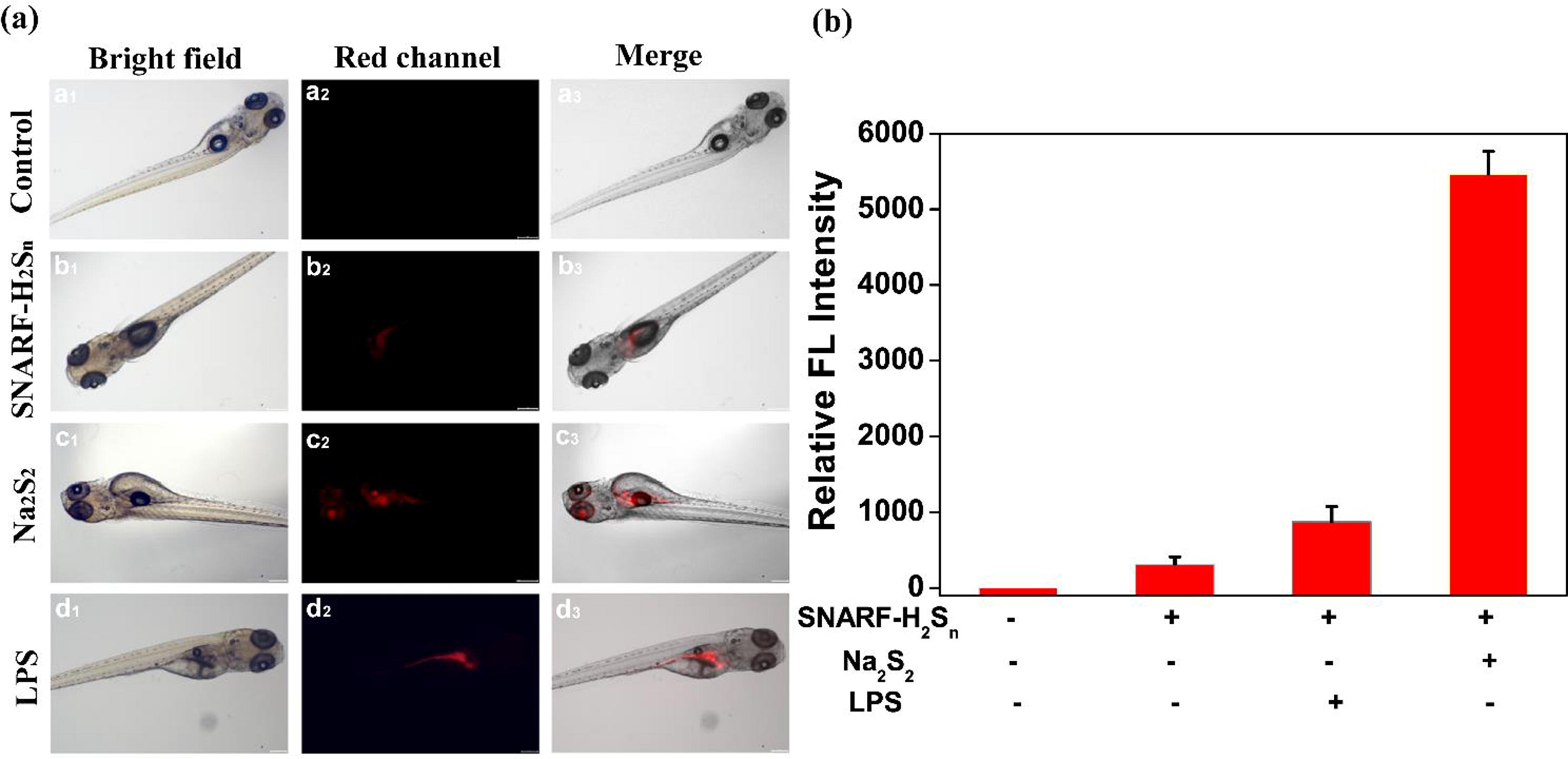
Fig. 3. Fluorescence imaging of exogenous/endogenous H2Sn in zebrafish. (a1–a3) control group; (b1–b3) the zebrafish were stained with 10 μM SNARF-H2Sn for 30 min; (c1–c3) the zebrafish were stained with 50 μM Na2S2 for 1 h, and further incubated with 10 μM SNARF-H2Sn for 30 min; (d1–d3) the zebrafish stimulated with LPS for 12 h, and then incubated with 10 μM SNARF-H2Sn for 30 min. (a1, b1, c1, d1) bright field images; (a2, b2, c2, d2) red channel images; (a3, b3, c3, d3) merged images. Scale bar = 200 μM. (b) Relative fluorescence intensity of red channel in panel A. Values represent mean standard error (n = 3).
Conclusion
In this work, by introducing phenyl 2-(benzoylthio) benzoate as a H2Sn-active trigger to SNARF scaffold, we reported a new type of red-emitting fluorescent probe named SNARF-H2Sn allowed for detection of H2Sn. On basis of dual reactivity of H2Sn, SNARF-H2Sn was able to respond H2Sn with extreme high selectivity and nanomolar detection limit. Leveraging SNARF-H2Sn, we enabled to track the level fluctuations of exogenous H2Sn in living cells and zebrafish by fluorescence microscope. What’s more, our results further revealed that SNARF-H2Sn successfully realized the imaging of endogenous H2Sn stimulated by LPS in vitro and in vivo. We anticipated that SNARF-H2Sn might be a valuable tool for researchers to reveal more information about H2Sn-related diseases.
https://blog.sciencenet.cn/blog-2438823-1258407.html
上一篇:Analysis extracellular vesicles by theranostic nanoplatforms
下一篇:Microfluidics-Based Sensing of Biospecies
全部作者的其他最新博文
- • Triphenylamine-AIE Materials for Cancer Theranostics
- • Fluorescent Probe forRatiometric Monitoring of Peroxynitrite
- • Fluorescence Probe for Pathological Stages of Wound Healing
- • Macrophage M2 polarization to neurological damage
- • SERS-RCA biosensor for profiling dual miRNAs
- • a glutathione-activated near-infrared fluorescent probe